Research

Publications related to the work explained on this page can be found in the publications section below. Other work, not related to this page, can be found on Prof. Vandersickel's google scholar profile .

1. A unified topological theory of cardiac arrhythmia

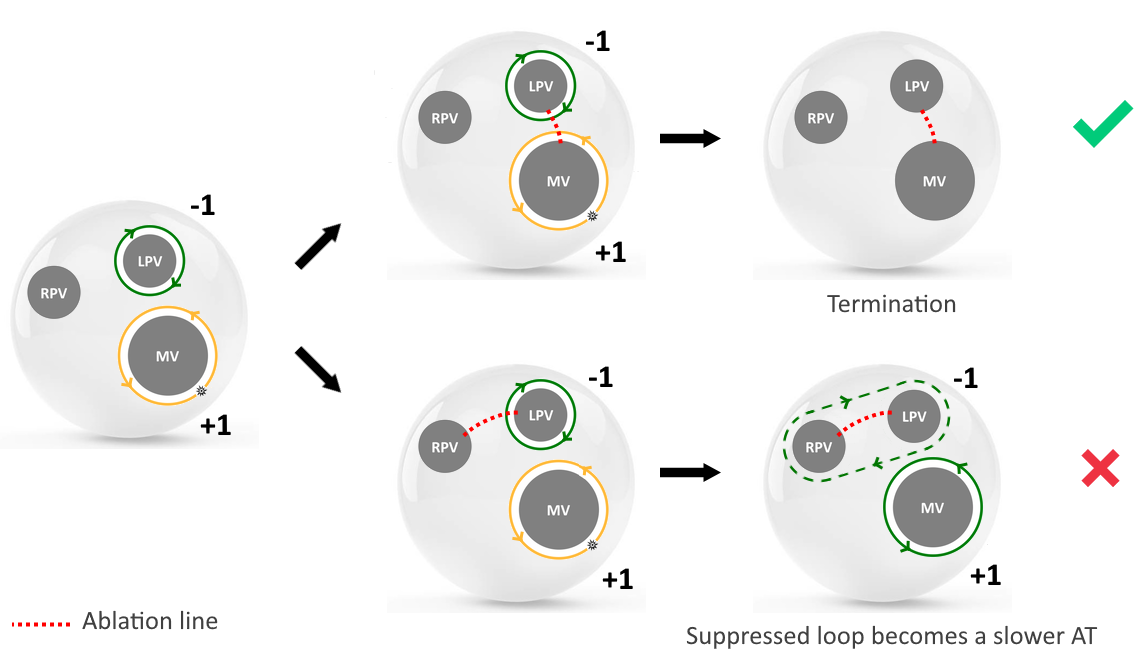
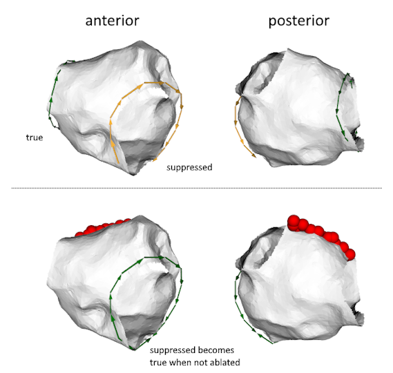
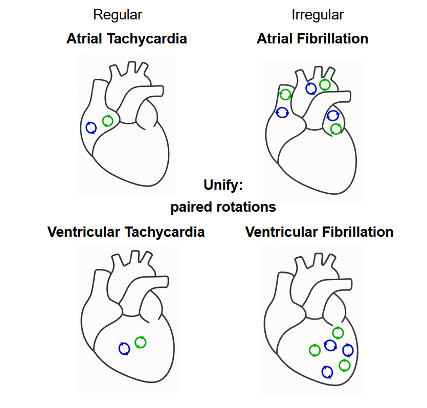
We hypothesize that reentry circuits generally do not exist as isolated entities but usually appear as paired objects: a clockwise reentry is inevitably accompanied by a counterclockwise reentry.

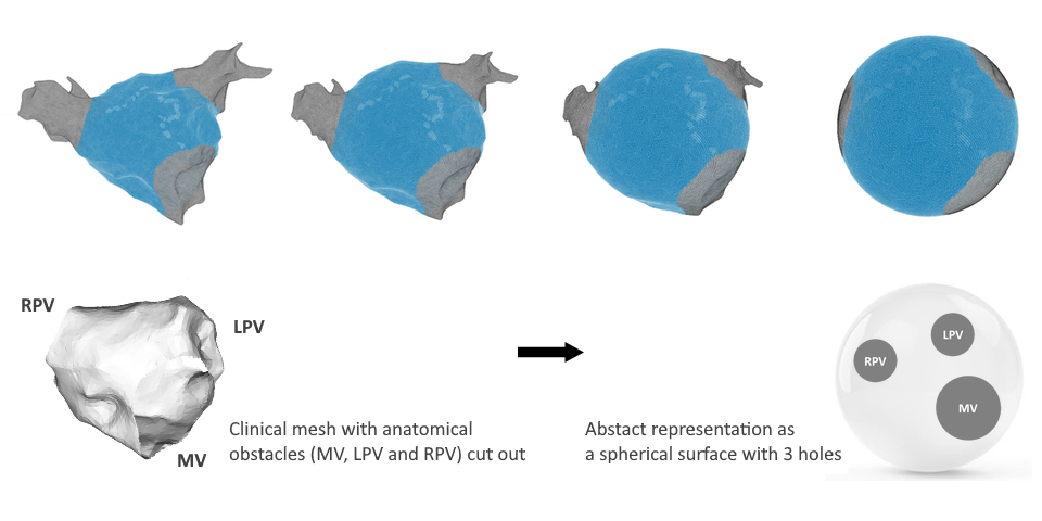
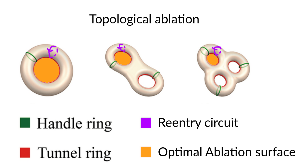
This conceptual breakthrough can be most easily understood by adopting a topological perspective. Topology is a branch of mathematics that studies the properties of objects that are invariant under stretch or deformation. It offers a useful way to simplify the heart’s structure without changing its key features. Topologically, the left atrium (LA) and right atrium (RA) can each be approximated as closed surfaces, equivalent to spheres with a finite number of boundaries10. The left ventricle (LV) and the right ventricle (RV) can also be described as spheres with boundaries, but with finite wall thickness, making their topology considerably more complex 11.

Atrium to sphere animation Topological transformation of a left atria into a sphere
Ventricle to sphere Animation Topological transformation of a left ventricle into a sphere with thickness

A largely forgotten topological theorem, the index theorem for phase singularities. The index theorem for phase singularities, first published in 1977, states that for phase fields defined on closed surfaces with boundaries, the sum of the topological indices must equal zero (Glass, 1977) (Davidsen et al, 2004). The cardiac electrical field serves as a prime example of such a phase field.

As we will show in the next sections, this simple theorem will unify all reentry-driven cardiac arrhythmia.

Unification of all major arrhythmia

2. OpenDGM: An open source package to analyze intra-cardiac data.

openDGM2,5 stands as a versatile tool capable of analyzing various arrhythmias, including Atrial Tachycardia (AT), Ventricular Tachycardia (VT), Atrial Fibrillation (AF), Ventricular Fibrillation (VF), and Torsade de Pointes (TdP). It efficiently processes files containing spatial coordinates and simultaneously measured intra-cardiac signals or local activation times (LATs) from electrodes.

While initially developed around network-based analyses, openDGM has evolved into a flexible platform that integrates multiple approaches, including advanced phase mapping, network theory, and topology, for comprehensive dataset analysis. Since becoming open source, we enabled the publication of complete analysis pipelines alongside scientific papers.

No regularity in spatial dataset locations is required, making openDGM independent of the measuring system. It accepts input from computational, experimental (such as needle or socket data), or clinical datasets (CARTO, RHYTHMIA, grid electrode, basket catheter data).

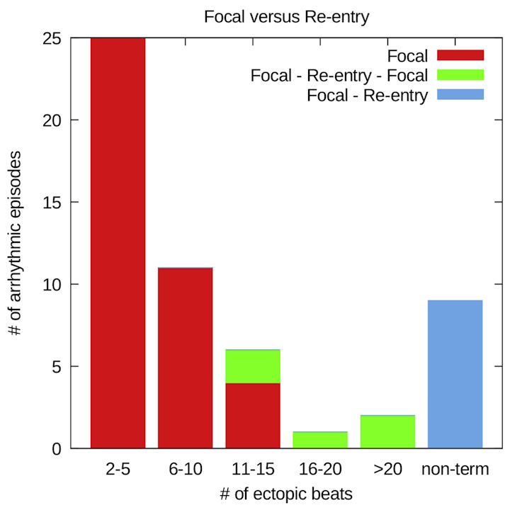
openDGM is recognized for its capability in analyzing reentry loops within arrhythmias, including complete reentry loops, but also incomplete reentry loops. Additionally, DGM can also identify focal sources, although we have not invested yet a large amount of research in this feature.

Mainly, our software package has implemented two main methods: network theory and phase mapping
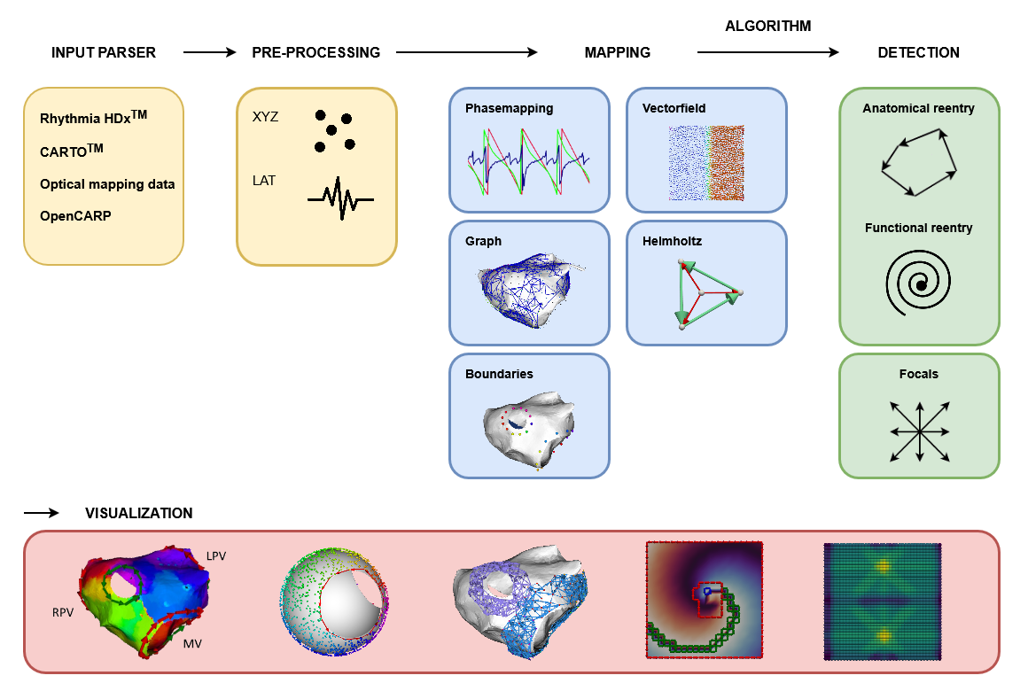
Network Theory

OpenDGM contains a method that transforms electrical mapping data from the heart into a directed network, making it possible to automatically detect the mechanisms driving an arrhythmia, we call this method Directed Graph Mapping (DGM).

DGM takes two simple inputs: the positions of the recording electrodes and their local activation times (LATs): the moment each electrode detects electrical activity. From these, a network is built where each electrode becomes a node, and arrows between neighbouring electrodes are drawn based on the direction and speed of electrical conduction. Only connections with physiologically plausible conduction velocities are included, ensuring the network faithfully represents true cardiac activation.

Once the network is constructed, DGM automatically searches for the arrhythmia source:

  • Reentrant activity is detected by finding directed cycles in the network, closed loops of electrical propagation;
  • Focal activity is identified by locating regions where all arrows point outward, indicating a local firing site.

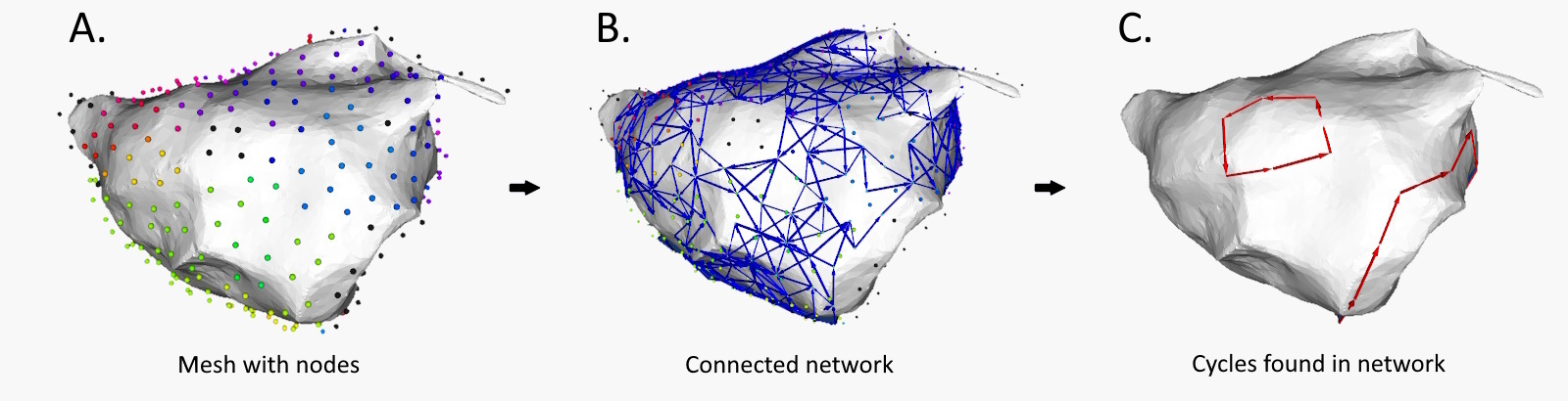
Extracting cycles from the network aligns with the identified reentry loop.

Phase Mapping

Phase mapping is one of the most widely used methods for detecting rotational drivers in cardiac arrhythmias. By converting electrical signals into a continuous phase representation, one can identify phase singularities: points around which rotational activity organizes. We have implemented two phase mapping methods in openDGM.

Naive phase mapping is the classical, widely-used approach. It assigns a phase value to each recording point for each time step and identifies rotors by detecting phase singularities. While powerful, this approach has a fundamental assumption: that the phase map is continuous across the entire tissue, except for a finite number of isolated points. In reality, cardiac tissue can contain conduction block, fibrosis, or anatomical boundaries, regions where the phase is either undefined or discontinuous. These regions, called phase defects, cause the naive approach to generate both false positive detections (rotational drivers that aren’t there) and false negatives (rotational drivers that are missed).

Extended phase mapping is an improved implementation developed by our group that explicitly detects and accounts for phase defects 18. Rather than assuming continuity, it identifies where the phase map breaks down, and correctly computes the phase index around these defects. This makes rotor detection robust in realistic tissue, including fibrotic substrates, anatomical boundaries, and clinical data, where the naive approach struggles. Across simulated, experimental, and clinical datasets, the extended approach eliminates erroneous detections and resolves previously missed rotational drivers. This has been overlooked for the past 30 years!

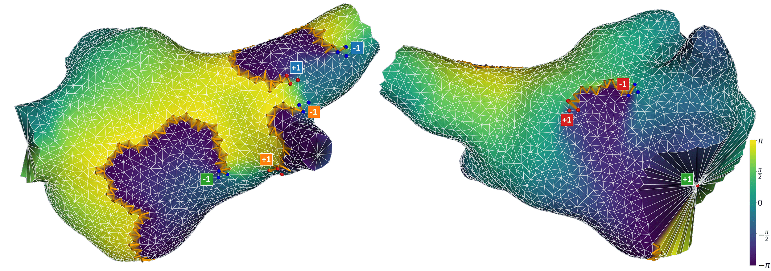
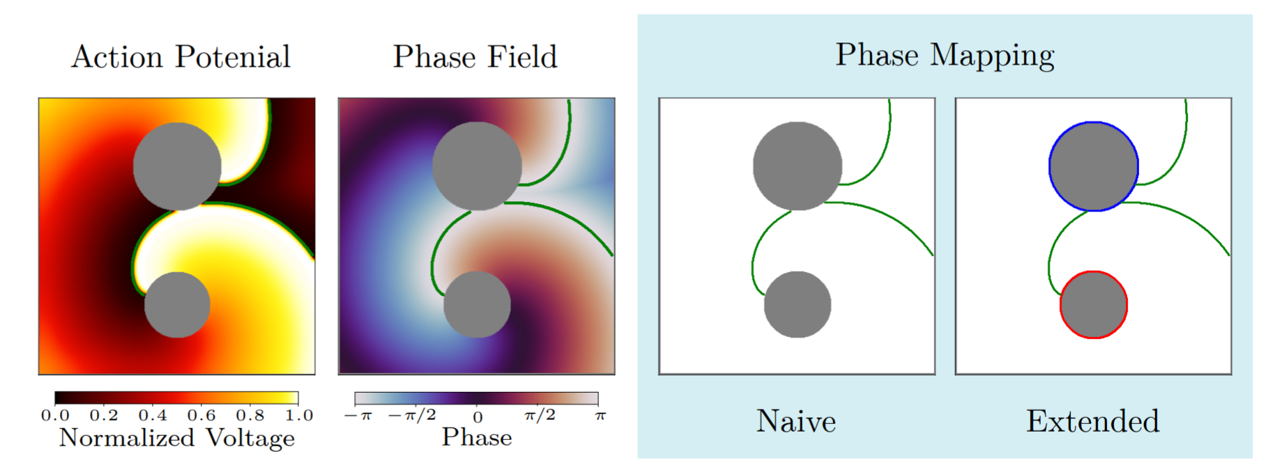
The naïve approach detects no phase singularities, as the phase within scar tissue is undefined. The extended approach instead identifies an average of two critical phase defects per time frame, with indices of +1 and −1 respectively.

Topology

OpenDGM also has algorithms that compute the index of given boundaries, for more on this see DGM-TOP

3. Research on Cardiac Arrhythmia

Below you can find an overview of our past and current studies about AT, AF, VT, VF, TdP, and some more general studies, often using (open)DGM for the analysis of the data.

Atrial Tachycardia · AT

Theoretical concept: Reentries come in pairs of two

Reentry is an underlying mechanism in many cases of atrial tachycardia (AT). While the prevalent belief for many years was that AT is mostly driven by a single reentry loop, we showed that this is usually not the case. The key factor behind this lies in the index theorem, prompting a deeper exploration into the atrial topology for a more nuanced understanding.

The left atrium (LA) and right atrium (RA) can each be topologically represented as a sphere with boundaries. The LA has at least three natural openings, the mitral valve (MV), left pulmonary veins (LPV), and right pulmonary veins (RPV), and the RA similarly features the tricuspid valve, inferior vena cava, and superior vena cava. Non-conductive scar tissue can act as additional boundaries, further shaping the atrial topology. Each patient's unique topology can therefore be characterized simply by counting the number of boundaries present.

It is crucial to note that ablation lines or natural lines of block connecting two boundaries effectively reduce them to a single topological boundary.

Transformation of left atrium to sphere

The index theorem states that topological charges on a closed surface must sum to zero (Davidsen et al, 2004) . For AT, this means reentrant loops always come in pairs.

While one reentry loop always completes a full rotation, its paired loop may be incomplete or suppressed due to collision with the first. Despite appearing less obvious, calculating the topological index around its boundary still yields ±1 — confirming it is equally relevant for treatment. We will can the boundaries with non-zero index the critical boundaries (CBs).

In an atrium with 2 boundaries due to previous ablation line(s), paired rotation essentially mirrors the clockwise and counterclockwise projection of a single reentrant circuit. When 3 boundaries or more are present, paired rotation indicates two clinically relevant loops around two boundaries (index +1, -1) with fused activation (index 0) encircling the other boundaries.

The direct termination of tachycardia can only be achieved by ablating the two boundaries with + and – indices. Failure to ablate the incomplete loop will result in the emergence of a slower AT. This explains why slower ATs often emerge after the first ablation line.

Ablation strategy

Each AT can be uniquely categorized by the number of boundaries and their topological index.

We validated our theory using approximately 600 simulations on spherical geometries with varying numbers (2–4) and sizes of boundaries, all of which were 100\% consistent with our hypotheses.

Our clinical work on AT is mainly performed in collaboration with Prof. Mattias Duytschaever and Prof. Sebastian Knecht from the hospital AZ Sint-Jan, Bruges, Belgium, who work with the CARTO system (Biosense Webster). We have analyzed over 131 AT cases retrospectively9,10, and 88 cases prospectively16,, supporting our hypothesis.

The spatial distribution of critical (CB) and noncritical boundaries (NCB) in 131 clinical atrial tachycardia cases analyzed retrospectively10,.
Automated Detection of Critical Boundaries after manual cut-out of all boundaries: DGM-TOP

We extended DGM with a new algorithm, DGM-TOP, capable of detecting both complete and incomplete reentry loops. DGM-TOP requires a correct manual cut-out of the boundaries in the input file, after which it calculates the topological index of each boundary and recommends the optimal ablation line for treatment.

Complete and Incomplete reentry
Automated Detection of Critical Boundaries without any manual intervention

Our extended phase mapping algorithm can also be applied to clinical AT case. By setting a threshold on the local phase gradient, we can accurately identify discontinuous regions in the atrial map.

Each discontinuity is a candidate for a re-entry circuit as rotational activity can only be present around phase discontinuities. By calculating the topological charge around the boundary of the discontinuity, we can find the critical and non critical boundaries.

An animation showing a suppressed re-entry circuit around an anterior scar and a complete re-entry around the mitrial valve. An animation showing a suppressed re-entry circuit around a scar and a complete re-entry around the mitral valve. The ablation line connecting these re-entries is shown by the red spheres.

The value of this method has already been proven as it aided in discovering a unique case of AT featuring a double figure-of-eight re-entry, where four re-entry circuits sustained the AT. 17

We are actively working on making this method more robust in a clinical setting.

Clinical AT (cycle length 280ms) with double figure-of-eight reentry. A–D: Activation maps. E: Topological scheme showing 4 CBs with a net charge of zero. F: The ablation strategy eliminates all 4 circuit loops; a lesion on the opposite side of the line of block would leave a viable circuit intact.

To aid development and help with analyzing clinical cases, we made a custom graphical interface where we can load in a clinical case and execute our algorithms on it. Using this we can immediately show the detected re-entry circuit. The detection method for phase defects and the threshold used can also be changed in the GUI. Ablation lines can be shown to check the detected re-entry circuits. For further analysis, we can also show the catheter points and EGMs.

Ventricular Tachycardia · VT

DGM can find complete reentry loops in VT on epi or endocardial maps

We tested an early version of DGM on VT mapped with CARTO (Biosense Webster), in collaboration with Associate Professor and Electrophysiologist Geoff Lee and Electrophysiology Fellow Dr. Josh Hawson from the Royal Melbourne Hospital7. Across 35 fully mapped VT procedures, 4 epicardial and 31 endocardial, DGM successfully identified complete reentry loops. At the time, however, we had not yet established that reentries always occur in pairs.

Ischemic cardiomyopathy following anteroseptal infarction. (A) Twelve-lead ECG of clinical VT with electrograms a–g (locations labelled in C). (B) Bipolar voltage map showing large anteroseptal scar. (C) LAT map illustrating dual-loop reentry (white arrow); a missing circuit segment >12.5% between blue and purple isochrones suggests an intramural component (black interrupted arrow), with a remote bystander channel (BC) showing diastolic activation. (D) ACVM incorrectly suggesting a focal mechanism due to centrifugal activation from an early activation focus. (E) DGM correctly identifying dual reentry with isthmus at the anterior septum.
Four Topologies of Paired Reentry in Ventricular Tachycardia

However, we later found that just as in AT, also for VT reentries have to obey the index theorem. However, as the ventrical wall has a thickness, each layer of the wall should obey the index theorem. Let us assume that reentries are anchored at scar tissue.

In this case, the shape of the underlying scar gives rise to four distinct paired reentry patterns: I-shaped, O-shaped, and U-shaped, each with specific implications for ablation. Successful ablation should connect each critical boundary pair, a strategy that coincides with targeting the protected VT isthmus11. We generated simulations for each scenario, where index calculations and virtual ablation experiments consistently confirmed our hypothesis.

Ventricle Critical Boundaries A representation of the four re-entry patterns in VT along with their optimal ablation.
From Boundaries to Tori: A Unified Topological Framework for Ventricular Tachycardia Ablation

In a next step, we have found a new topological framework for identifying optimal ablation targets in ventricular tachycardia (VT). Our previous IOU model classified CBs of cardiac tissue by detecting CBs on every tissue layer. However, this model could not generalize for realistic three-dimensional geometries due to the complexity of analyzing infinitely many tissue layers. To overcome this, this study reframes the problem using concepts from algebraic topology, modeling the heart and its pathological structures (such as scar and refractory tissue) as obstacles and boundaries embedded in three dimensions.

A central insight comes from the application of the index theorem in 3D, which constrains how electrical activity can circulate along boundaries. This leads to the key result that all critical boundaries sustaining VT must form closed surfaces with the topology of a torus. In this framework, previously distinct boundary types (I-, O-, and U-types) are unified as toroidal structures.

A ventricle scar will be topologically equal to an object with a certain genus.

These torus-shaped boundaries contain “handles” and “tunnels,” which correspond to fundamental topological features. The study shows that VT circuits are associated with specific “critical” handles and tunnels, and that these structures can be systematically identified using tools like the shortest homology basis.

Shortest homology basis.

Building on this theory, the concept of “topological ablation” is proposed: instead of targeting tissue empirically, clinicians can identify and ablate regions corresponding to critical tunnels, effectively disrupting the reentrant circuits that sustain VT. The approach is general and applies to arbitrary scar geometries, offering a principled method to locate ablation targets in 3D. Clinically, this suggests combining imaging data (such as MRI for scar tissue) with limited activation maps to pinpoint likely critical regions, potentially improving the precision and success rate of VT treatments while reducing unnecessary tissue damage.

Atrial Fibrillation · AF

We further demonstrated that the index theorem extends to irregular, time-varying reentries in computational models relevant to AF, indicating that paired reentries persist even under highly complex dynamics12. Moreover, we showed that AF can only be terminated by connecting reentrant circuits of opposite chirality along the propagating wavefront. This hypothesis is supported by a set of 600 simulations. Whether these findings translate to the clinical setting remains an open question.

Paired reentries in a model of AF.

Ventricular Fibrillation · VF

Although we have not investigated VF ourselves, in (Pertsov et al., PRL, 2000) the authors found that scroll wave filaments must start and end on medium boundaries. Moreover, in a simulation study by (Clayton et al., Progress in Biophysics and Molecular Biology, 2006) the authors also only found I,O,U-type filaments in 3D VF simulations

Torsade de Pointes · TdP

The initial application of DGM (primitive version) focused on analyzing intramural needle data from the CAVB dog model developed in Prof. M. Vos's lab at Utrecht Medical Center. The primary question was whether TdP is perpetuated by focal activity or reentry. Preliminary analysis using an early version of DGM revealed that short TdP episodes are sustained by focal activity, while longer episodes involve reentry. Non-terminating TdPs consistently showed reentry. The findings of this research have been published in 2017 in JACC: Clinical Electrophysiology.1.

After the results were published, an editoral was written on our work by Sachin Nayyar, Andreu Porta-Sánchez, and Kumaraswamy Nanthakumar (JACC EP). The question was raised how the reentry exactly manifests itself. Is it functional or anatomical? Are there multiple reentries? Is there meandering of the spiral core. This editorial inspired a follow-up paper where we sought to address some of these questions.

In our follow-up study6, we used the more mature DGM software, which confirmed our previous results. In addition, we found that non-terminating (NT) Torsade de Pointes (TdP) episodes consistently exhibited more simultaneous reentry loops compared to self-terminating (ST) episodes. (Bi-)ventricular loops were prevalent in non-terminating episodes. Thus we found that macro-reentry and multiple simultaneous localized reentries play a role in prolonged TdP. We also found that focal sources (which initiate each episode of TdP) tended to occur in preferred locations, suggesting potential implications for targeted treatment.

DGM (network algorithm) and Phase mapping

In our first study, we have compared DGM (thus using network theory) with phase mapping for a simulated stable rotor (functional reentry) whereby we put an regular electrode system on top of the rotor. We systematically added Gaussian noise to the LATs, and compares DGM with phase mapping. We found that phase mapping is very sensitive to noise, while DGM is much more robust.

In collaboration with Prof. Saiz and Dr. Martínez Mateu we have also compared phase mapping and DGM for the analysis of simulated basket catheter data in the right atrium for a meandering spiral. We found that DGM overcomes some of the limitations of phase mapping, which often finds false rotors4.

Finally, we conducted a comparative analysis of various phase mapping techniques alongside DGM for rotor detection, evaluating performance using 64 simulated 2D rotors with varying levels of fibrotic tissue, temporal noise, and meandering. Our study indicates that under conditions of low meandering, fibrosis, and noise, both PM and DGM yield comparable, excellent results. However, under conditions of high meandering, fibrosis, and noise, phase mapping is prone to errors, particularly an excess of false positives, leading to lower precision. In contrast, DGM demonstrates greater robustness in these scenarios8.

Publications